
News | TCTAP 2024
AI Application of Cardiovascular Imaging
MedTech Innovation
Jihoon Kweon
Asan Medical Center, Republic of Korea
A recent lecture at TCTAP 2024 introduced a new database for developing cardiovascular imaging artificial intelligence (AI), integrating AI models for post-percutaneous coronary intervention (PCI) evaluation, and application of general AI.
Jihoon Kweon, PhD (Asan Medical Center, Seoul, Korea) has developed AI-QCA, which provides reproducible and reliable real-time coronary angiography analysis. To achieve this, data from 10,000 patients were used to train the AI, minimizing exclusions better to reflect the characteristics of the real patient pool. However, the challenge of expanding the database size to develop a more robust AI model remained. To address this, he built a large-scale database (Asan Invasive Coronary Angiography Network, A-ICAnet) containing 3 million angiographic frames, with plans to increase this to 100 million images (Figure 1). He demonstrated that by using A-ICAnet, segmentation of target vessels could be improved by more than 2% on average. He also suggested that A-ICAnet could be used for segmenting lumens in other views or with different interventional devices.
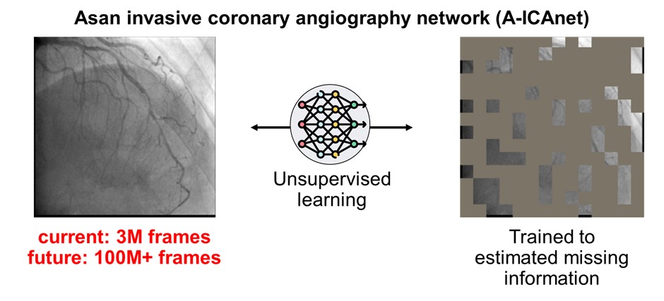
Figure 1. A large-scale coronary angiography database for AI development
AI integration was then introduced to post-PCI stent evaluation. Although stent expansion can be evaluated by specifying the proximal and distal ends of the implanted stent using AI-QCA, eliminating manual operations will help incorporate post-PCI stent evaluation into clinical practice. He developed an AI model for stent segmentation in fluoroscopy to automatically set the reference points for post-PCI stent evaluation. Over 1,000 images were collected at Asan Medical Center and used for AI training. Using an ensemble of three segmentation models, the AI achieved an F1 score of over 88%. By mapping the stent area onto the angiography, the proximal and distal ends of the stent area could be determined accurately (Figure 2). Post-PCI evaluation using AI-QCA was completed within one second without additional image acquisition. In the representative case, the AI model accurately set the stent area for assessment and identified an under-expansion that would likely be difficult to estimate visually.
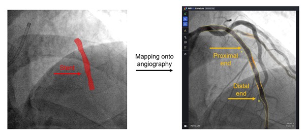
Figure 2. AI-AI integration for post-PCI stent evaluation
Lastly, he demonstrated the potential of general AI for cardiovascular imaging. Meta has developed an AI model called the Segment Anything Model (SAM), which segments each area based on user input. With SAM, a catheter or guidewire could be automatically segmented with just one click (Figure 3). Although this task previously required advanced image processing methods, it now requires a single AI model without labeling, training, or tuning. Meta's Co-tracker was developed to track the path at a given location. By locating the stent area in fluoroscopy, the Co-tracker could continue to track movement even after the lumen area was filled with contrast. He claimed that while these technologies are not yet sufficient to analyze all cardiovascular imaging, AI advancements are expected to change the clinical environment in the near future.
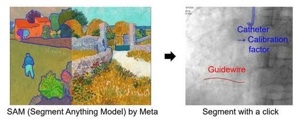
Figure 3. Application of general AI for segmentation of fluoroscopy
Hot Topics
MedTech Innovation
Friday, April 26, 2:00 PM ~ 3:08 PM
Presentation Room 1, Level 1
Edited by
Jihoon Kweon, MD
Asan Medical Center, Korea (Republic of)
Jihoon Kweon
Asan Medical Center, Republic of Korea
A recent lecture at TCTAP 2024 introduced a new database for developing cardiovascular imaging artificial intelligence (AI), integrating AI models for post-percutaneous coronary intervention (PCI) evaluation, and application of general AI.
Jihoon Kweon, PhD (Asan Medical Center, Seoul, Korea) has developed AI-QCA, which provides reproducible and reliable real-time coronary angiography analysis. To achieve this, data from 10,000 patients were used to train the AI, minimizing exclusions better to reflect the characteristics of the real patient pool. However, the challenge of expanding the database size to develop a more robust AI model remained. To address this, he built a large-scale database (Asan Invasive Coronary Angiography Network, A-ICAnet) containing 3 million angiographic frames, with plans to increase this to 100 million images (Figure 1). He demonstrated that by using A-ICAnet, segmentation of target vessels could be improved by more than 2% on average. He also suggested that A-ICAnet could be used for segmenting lumens in other views or with different interventional devices.
AI integration was then introduced to post-PCI stent evaluation. Although stent expansion can be evaluated by specifying the proximal and distal ends of the implanted stent using AI-QCA, eliminating manual operations will help incorporate post-PCI stent evaluation into clinical practice. He developed an AI model for stent segmentation in fluoroscopy to automatically set the reference points for post-PCI stent evaluation. Over 1,000 images were collected at Asan Medical Center and used for AI training. Using an ensemble of three segmentation models, the AI achieved an F1 score of over 88%. By mapping the stent area onto the angiography, the proximal and distal ends of the stent area could be determined accurately (Figure 2). Post-PCI evaluation using AI-QCA was completed within one second without additional image acquisition. In the representative case, the AI model accurately set the stent area for assessment and identified an under-expansion that would likely be difficult to estimate visually.
Lastly, he demonstrated the potential of general AI for cardiovascular imaging. Meta has developed an AI model called the Segment Anything Model (SAM), which segments each area based on user input. With SAM, a catheter or guidewire could be automatically segmented with just one click (Figure 3). Although this task previously required advanced image processing methods, it now requires a single AI model without labeling, training, or tuning. Meta's Co-tracker was developed to track the path at a given location. By locating the stent area in fluoroscopy, the Co-tracker could continue to track movement even after the lumen area was filled with contrast. He claimed that while these technologies are not yet sufficient to analyze all cardiovascular imaging, AI advancements are expected to change the clinical environment in the near future.
Hot Topics
MedTech Innovation
Friday, April 26, 2:00 PM ~ 3:08 PM
Presentation Room 1, Level 1
Edited by
Jihoon Kweon, MD
Asan Medical Center, Korea (Republic of)
Leave a comment